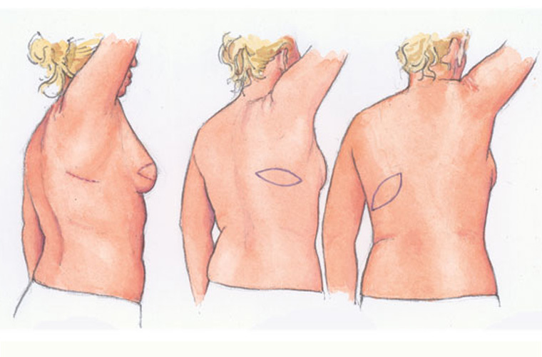
Latissimus Dorsi Breast Reconstruction uses muscle from the patients back, the blood vessels attached to the muscle and the skin overlying it.
The tissue which is moved is called a flap. You have a right and a left Latissimus Dorsi muscle. Each is a large, flat triangular muscle situated in the upper back, either side of your spine and normally helps to move your shoulder.
Despite the fact that part of the muscle is moved to your chest to make a new breast, your shoulder movement should not be affected in the long term. The remainder of the Latissimus Dorsi muscle will strengthen to compensate and you have other shoulder muscles which carry out the same function.
To give your breast more volume your surgeon may place a tissue expander or breast implant underneath the flap. If appropriate this will have been discussed with you prior to your operation.
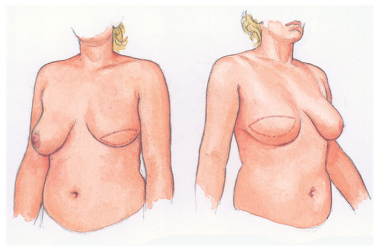
The paddle of skin attached to the underlying latissimus dorsi back muscle can be orientated at different angles depending on the tissue available, leaving a thin scar which maybe horizontal or lying along the line of the rib. The muscle is swung around underneath the armpit tissue and moved to the pocket within the breast to provide new skin for the breast reconstruction, but also to help protect the implant from the surgical scar while it is healing. The second diagram shows the skin from the back after it has been inset into the front portion of the reconstructed breast.
This second diagram shows the skin from the back after it has been inset into the front portion of the reconstructed breast.
Illustrations – David Hurrell
Other Procedures
Please go through the information below in your own time to help your understanding of the options available.
Meetings are held on Tuesday afternoons either virtually via Microsoft Teams video conferencing or face to face at St Andrews Hospital outpatients department.